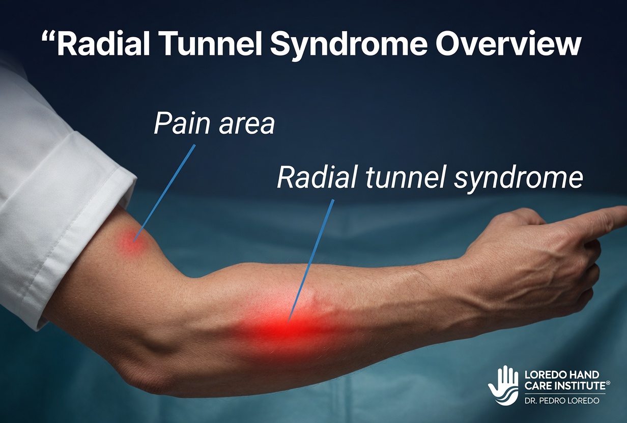
Radial tunnel syndrome is a deep aching pain over the upper outer forearm caused by compression of the posterior interosseous nerve (PIN) as it passes through the radial tunnel and dives beneath the supinator muscle at the Arcade of Frohse. The condition often coexists with, or is misdiagnosed as, tennis elbow. Patients typically have no measurable weakness on examination but experience pain that worsens with manual work, gripping, and forearm rotation. Treatment ranges from activity modification, splinting, and selective injection to surgical decompression of the nerve.
The Anatomy of the Radial Tunnel
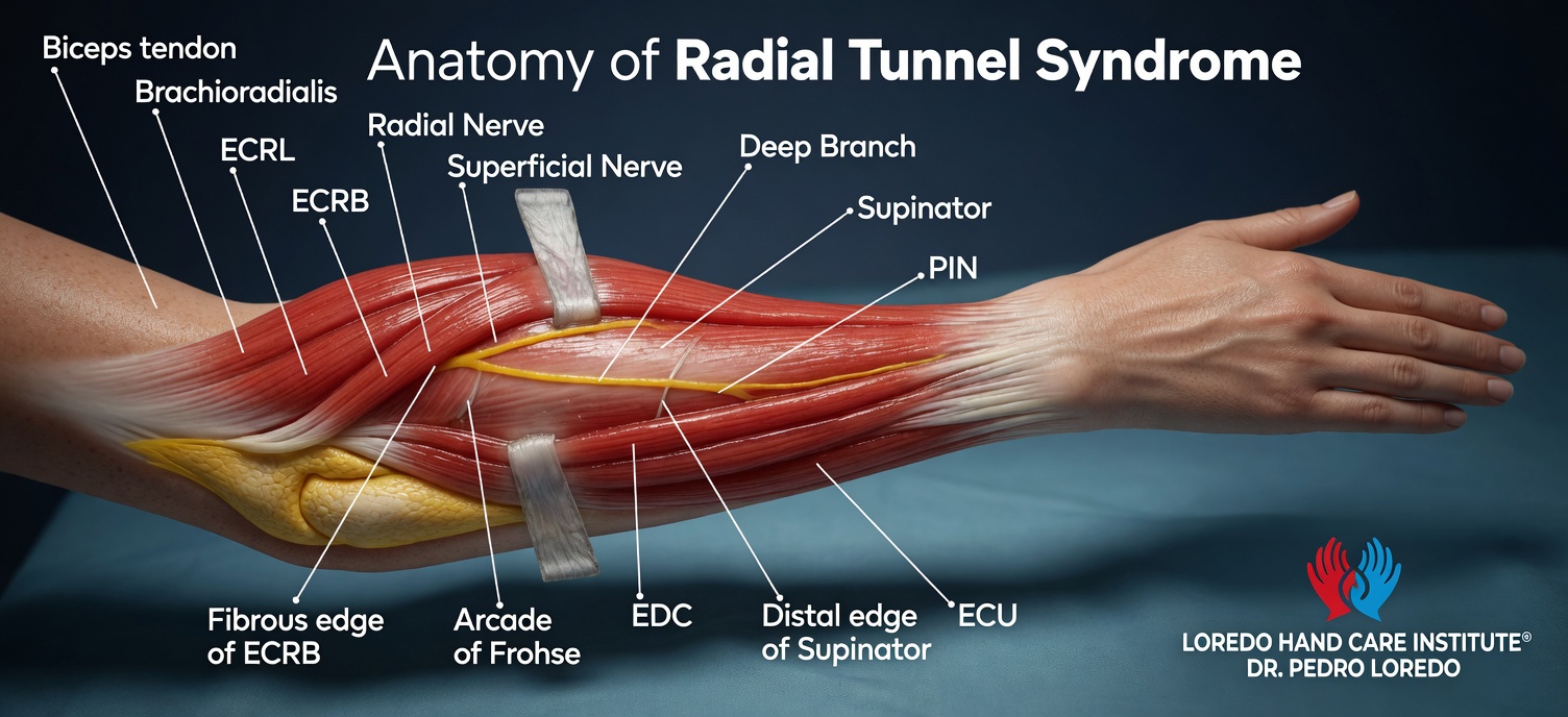
The radial nerve crosses the lateral elbow and divides into two branches: a superficial sensory branch that travels distally beneath the brachioradialis to supply sensation to the back of the thumb, and a deep motor branch called the posterior interosseous nerve (PIN). The PIN is the branch responsible for radial tunnel syndrome.
The radial tunnel is the anatomic space the PIN traverses on its way from the lateral elbow into the deep posterior compartment of the forearm. The tunnel is approximately 5 cm long. There are five potential compression points along the tunnel: fibrous bands at the radiocapitellar joint, the leash of Henry (a small group of recurrent radial vessels), the medial edge of the extensor carpi radialis brevis, the proximal edge of the supinator (the Arcade of Frohse), and the distal edge of the supinator. The Arcade of Frohse is the most common compression site and is the structure released during surgery.
Radial tunnel syndrome and posterior interosseous nerve syndrome are on a spectrum of the same nerve compression but present very differently. Radial tunnel syndrome is the painful compression variant: pain without weakness. PIN syndrome is the motor compression variant: weakness without pain, with finger drop and difficulty extending the digits at the MCP joints. They share an anatomic compression but produce distinct clinical pictures.
Clinical coding: ICD-10 G56.30 (lesion of radial nerve, upper limb, unspecified). SNOMED CT 200283005.
Causes and Risk Factors
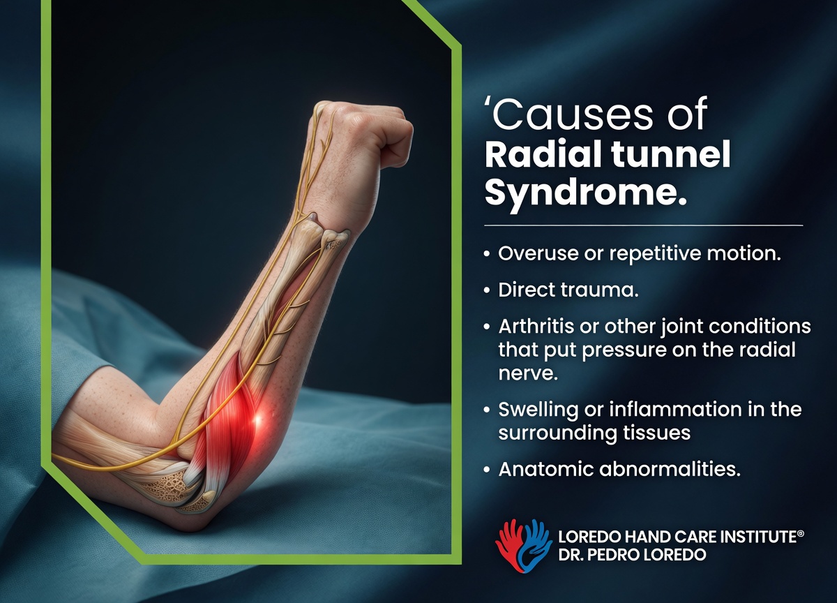
- Repetitive forearm rotation, particularly forced supination and pronation in manual work
- Forceful gripping with the elbow extended and forearm pronated
- Manual occupations: mechanics, butchers, assembly line workers, drywall installers
- Recreational activities: racquet sports with poor mechanics, weight training with heavy gripping
- Coexisting tennis elbow in 5 to 10 percent of patients
- Trauma to the proximal radius or supinator
- Anatomic variants such as a tendinous rather than muscular Arcade of Frohse
Symptoms and Warning Signs
- Deep aching pain over the upper outer forearm, 4 to 5 finger-breadths distal to the lateral epicondyle
- Pain that worsens with gripping, lifting, or sustained forearm rotation
- Pain that may radiate up to the elbow or down the forearm
- Symptoms that interfere with sleep due to a positional ache
- No measurable weakness on examination (a key feature)
- No numbness or tingling, distinguishing it from carpal tunnel and cubital tunnel
- Coexisting tennis elbow symptoms in some patients
- Pain reproduced by specific provocative tests rather than by general tenderness over a tendon
How the Diagnosis Is Made
Radial tunnel syndrome is a clinical diagnosis. Imaging and electrodiagnostic studies are often normal and are mainly used to exclude alternative diagnoses. Dr. Loredo performs three standard provocative tests:
- Middle finger extension test: with the elbow extended, the patient extends the middle finger against the examiner's resistance. Reproduction of pain over the radial tunnel (not at the lateral epicondyle) is positive.
- Resisted supination test: with the elbow extended, the patient supinates the forearm against resistance. Pain over the radial tunnel suggests compression of the PIN.
- Point tenderness over the radial tunnel: tenderness localizes 4 to 5 finger-breadths distal to the lateral epicondyle, deep in the muscle belly. This contrasts with tennis elbow tenderness, which is 1 to 2 cm distal to the epicondyle directly over the ECRB origin.
Imaging is mainly used to exclude alternative diagnoses. X-ray rules out radiocapitellar arthritis or a synovial fold. MRI can show muscle denervation in chronic cases. EMG and nerve conduction studies are commonly normal in radial tunnel syndrome because the compression is dynamic and pain-driven, not consistently producing measurable conduction slowing. Normal studies do not rule out the diagnosis. A diagnostic injection of local anesthetic into the radial tunnel can be useful in difficult cases.
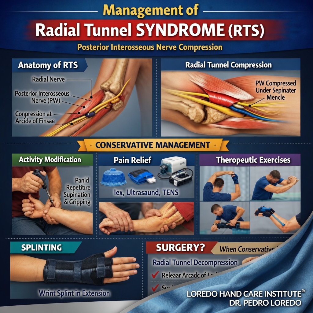
Non-Surgical Treatment Options
At least 3 to 6 months of structured conservative treatment is appropriate before considering surgery.
- Activity modification: avoid the forceful gripping and forearm rotation pattern that produces symptoms. Modify work tasks where possible.
- Splinting: a long-arm or wrist-elbow splint with the elbow at 90 degrees and the forearm in slight supination reduces nerve tension. Worn at night and during aggravating tasks.
- Targeted hand therapy with nerve gliding exercises, gentle stretching, and progressive forearm strengthening once acute pain settles.
- Topical and oral nonsteroidal anti-inflammatory medication for symptom relief.
- Selective corticosteroid injection into the radial tunnel under ultrasound guidance can be both diagnostic and therapeutic.
- Treatment of coexisting tennis elbow when present, since the two conditions reinforce each other.
Surgical Options
Surgery is considered when symptoms persist beyond 3 to 6 months of structured conservative treatment and when the patient is significantly limited in work or daily activity.
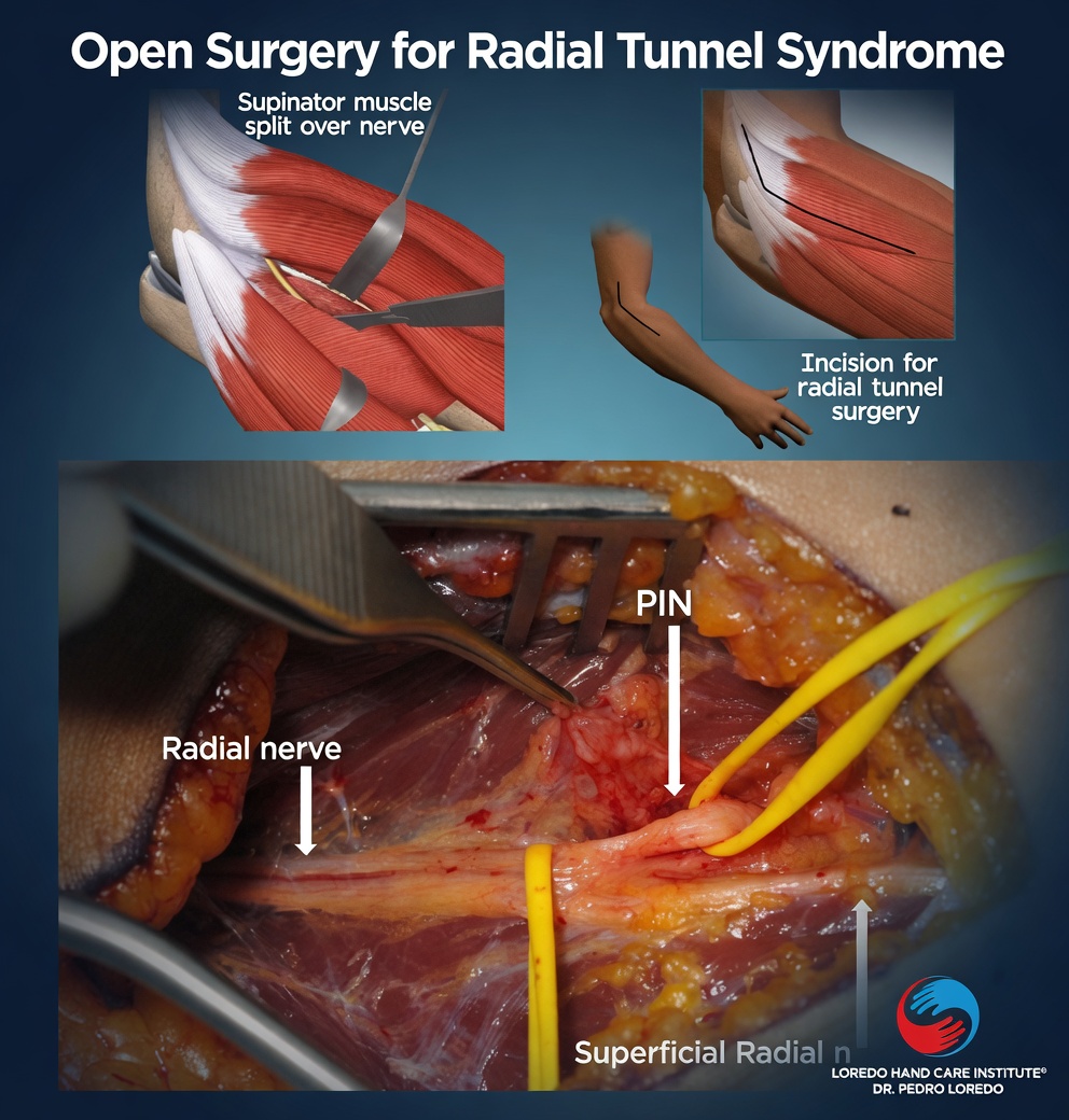
Posterior Interosseous Nerve Decompression
- Small incision on the lateral proximal forearm
- The brachioradialis muscle is retracted to expose the radial nerve and its branches
- The radial nerve is identified and traced through the tunnel
- All five potential compression points are released, with particular attention to the Arcade of Frohse at the entrance to the supinator
- The procedure takes 30 to 45 minutes under regional anesthesia
- Soft dressing and a removable splint for the first week
Combined Procedure with ECRB Release
When tennis elbow coexists with radial tunnel syndrome, both can be addressed in the same operation through related approaches. Combined surgery is appropriate when both conditions have been clinically established and conservative treatment has failed.
Surgical success rates for radial tunnel decompression are 60 to 80 percent across published series, lower than for other peripheral nerve decompressions. Patient selection is the most important variable. Workers' compensation cases and patients with chronic regional pain syndromes have somewhat lower success rates.
Recovery Timeline
- Day 0: Procedure performed in 30 to 45 minutes under regional anesthesia. Removable splint applied.
- Day 1 to 7: Light hand and wrist motion several times per day. Bandage care per instructions.
- Week 1 to 2: First follow-up. Sutures removed. Begin gentle elbow range of motion.
- Week 2 to 6: Progressive forearm range of motion and gentle strengthening. Hand therapy as needed.
- Week 6 to 12: Pain typically continues to improve over months. Return to manual labor and sport in this window.
- Month 3 to 6: Final results assessed at 6 months. Some patients continue to improve gradually beyond that point.
Returning to Work and Daily Activity
- Office or desk work: 1 to 2 weeks after surgery
- Light manual labor: 4 to 6 weeks
- Heavy manual labor: 8 to 12 weeks with attention to the technique that produced symptoms
- Racquet sports: 3 to 4 months with a coached return through stroke mechanics review
- Driving: 1 to 2 weeks once off pain medication and able to grip the wheel without sharp pain
Frequently Asked Questions
How is radial tunnel syndrome different from tennis elbow?
Both conditions cause pain on the outside of the elbow and forearm, and they often coexist. The differences are in the location of maximal tenderness and in the response to provocative tests. Tennis elbow tenderness localizes 1 to 2 cm distal to the lateral epicondyle, directly over the ECRB tendon origin. Radial tunnel tenderness sits 4 to 5 finger-breadths distal to the epicondyle, deep in the muscle belly over the supinator. Resisted middle finger extension and resisted supination reproduce radial tunnel pain more reliably than tennis elbow pain. Many patients with persistent or recurrent tennis elbow are eventually found to have an unrecognized radial tunnel component.
Will I have weakness with radial tunnel syndrome?
Usually no. Radial tunnel syndrome is the painful compression variant of posterior interosseous nerve compression and typically does not produce motor weakness. The closely related condition called posterior interosseous nerve syndrome produces drop fingers and weakness without pain. The two are on a spectrum of the same nerve compression but present very differently. Radial tunnel pain without weakness is the classic presentation.
Why are EMG and nerve studies often normal?
Radial tunnel syndrome is a clinical diagnosis. Nerve conduction studies and EMG can be normal because the compression is often dynamic and pain-driven rather than producing measurable nerve conduction slowing. Normal electrodiagnostic studies do not rule out the diagnosis when the clinical picture is consistent. Imaging is similarly limited and is mainly used to exclude alternative diagnoses.
How long should I try conservative treatment before considering surgery?
At least 3 to 6 months of activity modification, anti-inflammatory medication, splinting, and selective injection trials are appropriate before considering surgery. Some patients improve with simple changes in repetitive motion patterns. Surgery is reserved for patients who have completed a structured conservative course and continue to be limited in work or daily activity.
What does PIN decompression involve?
Posterior interosseous nerve decompression is an outpatient procedure performed through a small incision on the lateral forearm. The brachioradialis muscle is retracted, the radial nerve is identified and traced, and the deep branch (PIN) is followed into the supinator muscle. The Arcade of Frohse, a fibrous arch at the entrance to the supinator, is divided to relieve compression. Other potential compression points along the nerve are also released. The procedure takes 30 to 45 minutes under regional anesthesia. Recovery is similar to other elbow surgeries.
When to Call the Doctor
Before treatment, call our office for evaluation if you experience:
- Lateral elbow or upper outer forearm pain that has lasted more than 6 weeks
- Persistent or recurrent tennis elbow symptoms despite appropriate treatment
- Pain that worsens with gripping, lifting, or forearm rotation but without numbness or weakness
- Failure to improve with structured conservative care
Call promptly for any of the following, which suggest a different diagnosis:
- New finger drop or inability to extend the fingers (suggests true PIN syndrome with motor loss)
- Numbness or tingling in the hand (suggests carpal tunnel, cubital tunnel, or another nerve problem)
- Visible swelling, warmth, redness, or fever
- Pain after a fall or direct blow to the elbow or forearm
After surgery, call promptly if you experience fever over 101°F, expanding redness or drainage at the incision, severe pain not controlled by medication, or new numbness or weakness in the hand.
For any medical emergency, call 911 or go to the nearest emergency department.
Related Conditions
- Tennis Elbow (Lateral Epicondylitis): the most important alternative diagnosis. The two conditions can coexist and reinforce each other. Differentiated by the specific location of tenderness and response to provocative tests.
- Cubital Tunnel Syndrome: another nerve compression around the elbow, but on the medial side and producing numbness in the ring and small fingers. The differential is usually clear.
- Carpal Tunnel Syndrome: median nerve compression at the wrist. Listed because patients with persistent forearm pain sometimes have multiple coexisting nerve issues.
- Pronator Teres Syndrome: median nerve compression in the proximal forearm. The medial counterpart to radial tunnel syndrome on the same forearm rotation pattern.
- Hand and Wrist Arthritis: arthritis can refer pain into the forearm and rarely produce confusion with nerve compression diagnoses.
From the Blog
- Tennis Elbow vs Golfer Elbow: key differential diagnosis with lateral elbow pain.
Watch: Radial tunnel syndrome education
Short videos from Dr. Loredo's YouTube and Facebook channels.